
Radiation therapy, or "radiotherapy," is the treatment of cancer using ionizing radiation. Radiotherapy works by irradiating cancer cells, which harms and ultimately destroys them. For certain types of cancers, radiotherapy can be delivered by internal radioactive implants, which is called brachytherapy. However, many more types of cancer are treated by delivering the radiation using targeted beams from an external source called a linear accelerator. This is commonly referred to as external-beam radiation therapy (EBRT). This is what we use at USLV Radiation and Imaging Center (see more specifics below).
How does external beam radiation therapy work?
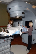
The linear accelerator creates the calculated dose of radiation, a beam which is typically only "on," for several minutes per treatment. The actual treatments are administered in a large vault-like room and are completely safe and do not cause you to become radioactive. Afterwards, it is totally safe for family members and friends to be around you. The beam of ionized radiation is directed at the cancerous tumor, affecting the cancer cells by disrupting their faulty DNA so they are no longer able to grow, and ultimately they degenerate and die. However, normal cells, in the proximity of the cancer, possess healthy DNA and have cellular mechanisms that allow their DNA to withstand the radiation's effect. Over the course of multiple treatments and time, the cancer is destroyed. Side effects result from the small percentage of normal cells in the treatment area which are affected by the radiation. Minimizing their exposure is one of the goals of modern techniques.
Treating the cancer with minimal exposure to normal tissue
Although normal cells have the ability to recover, the goal of radiation therapy is to maximize the dose to the cancer cells while minimizing exposure to normal tissue (and, therefore, the risks and severity of side effects). The challenge is to conform the radiation delivery as precisely as possible to the location, shape, size and orientation of the cancer. Over the years, EBRT has advanced to offer modalities that are ever-better at targeting and conformance in order to shrink and destroy tumors:
-
Three-dimensional conformal radiation therapy (3DCRT) – Using three-dimensional imaging (computed tomography, or "CAT scan"), our oncologists and physicists can devise a treatment based on—and customized to—a cancer's shape, size, location and proximity to other normal anatomic structures.
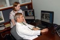
Treatment plan being devised - Intensity-modulated radiation therapy (IMRT) – Powerful computer software lets us plot treatment angles and varied beam intensities based on the true size, shape, density, location and orientation of the cancer. Pairs of tungsten "leaves" extend into and retract out of the radiation beam, shaping smaller beams and modulating radiation intensity for even greater 3D conformance. This effectively “wraps” the high dose region tightly around the target. Treatments are typically no more than a couple of minutes long, five days a week, for approximately eight or nine weeks for prostate cancer, and 3-8 weeks for other malignancies.
- RapidArc™ – an advanced form of IMRT, now routinely utilized by our radiation oncologists, Dr. Alden and Dr. Mehta at USLV, the RapidArc Linear Accelerator radiation therapy technology allows very complex treatments to be delivered in ten minutes or less, precisely to the targeted area, sparing healthy tissue, in contrast to the earlier technologies which required a 15- to 25-minute treatment to achieve the same results. RapidArc permits the entire radiation dose to be administered to the entire tumor with just one single rotation of the machine around the patient. This means that there is less chance of inadvertent patient or organ movement during treatment, so it reduces chances of having targeting errors, and potentially less side effects. Most importantly, RapidArc can achieve the same high cure rates for urologic cancers as the previous delivery systems.
- Image-guided radiation therapy (IGRT) – Because the cancer site (and normal structures) can move between planning and treatment and day to day, it is customary to expand the treatment region by a "margin" to allow for this movement. The advancement with IGRT, is that we use imaging technology to precisely locate the target site at the moment before each treatment, which improves accuracy, reduces the treatment margins and further minimizes exposure to normal tissue. Ultimately, this prevents excess high dose radiation exposure to your healthy tissue, and minimizes the chance of side effects.
External beam radiation therapy treatment planning
Simulation
After an initial consultation with a radiation oncologist and the decision has been made to proceed with EBRT, the next session is usually a planning session, which is called a simulation. During this session, the radiation treatment area is designated, and 3D images or your body are acquired for subsequent planning. The radiation oncologist is aided by one or more radiation technologists. Of all the visits to the radiation oncology facility, the simulation session may actually take the most time. During simulation, patients lay on a table somewhat similar to that used for a CT scan. The table can be raised and lowered and moved forward and back. The room is periodically darkened while the images are taken and the treatment fields are being set. Temporary marks may be made on the patient's skin with markers. The simulation may last anywhere from 25 minutes to an hour or more, depending on the complexity of what is being planned.
Once the aspects of the treatment fields are satisfactorily set, and the 3D CT images of your treatment area are taken, you will be given several tattoo dots marking the initial set up point. After that you may go home. Meanwhile, the radiation oncologist, in conjunction with a physicist and dosimetrist, will precisely map out your treatment plan specific to your individual anatomy and prescribe the dose and other treatment specifics. The plan will be thoroughly checked by your radiation oncologist and tested by the physicist. All plans are actually first, "treated live" by the linear accelerator on a test dummy-phantom to see that the dose and shape of the delivery is what your oncologist prescribed for you. This whole planning process takes about 5-7 days.
The treatments are instituted only after the radiation oncologist and technologists have rechecked and agree on the calculations and treatment field, and are thoroughly satisfied with the patient's specific treatment plan. Radiation treatments are then administered in a room separate from the simulation room.