
Once a solid renal tumor is found on kidney imaging, because of the high risk they represent a kidney cancer, surgical excision is the standard treatment. Small renal tumors can often be treated, without sacrificing the entire kidney, by removing only the tumor and a small part of the kidney. Many studies have proven that this technique, termed partial nephrectomy, has similar cure rates to removing the whole kidney, without losing significant renal function. In the past, these operations were performed through a large and painful flank or upper abdominal incision, with risk of significant bleeding routine. Also, recovery time from such an operation was lengthy and difficult. The da Vinci Robotic Partial Nephrectomy has revolutionized kidney surgery by minimizing bleeding, speeding recovery, and maximizing preservation of renal function.
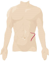
Open Left Kidney
Surgery Incision
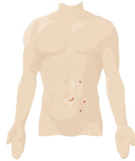
da Vinci Robotic Left Kidney
Surgery Port Incisions
Treating Kidney Cancer and da Vinci Robotic Partial Nephrectomy
Robotic laparoscopic partial nephrectomy with the da Vinci robotic system uses the robot and a minimally invasive approach (laparoscopy) to perform exactly the same procedure that is done in open or traditional laparoscopic approach. In any partial nephrectomy (open, laparoscopic, or robotic), the kidney cancer is removed with a small amount of normal tissue around it. The normal tissue around the cancer that is removed is known as the margin, and this tissue serves to assure that no cancer is left in the body. With the robotic laparoscopic procedure, no large incisions are required to perform the procedure. Instead, five or six 1/2 cm to 1 cm incisions (less than 1/2 inch) are made. The spaces in the body are gently filled with gas to make working space and a small camera is placed into the body through one of the incisions. The other small incisions are used to place working instruments, which can be used to perform the procedure.
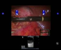
Ultrasound tumor identification
Usually, the kidney cancer can be readily seen using the da Vinci robot's optical system. If the cancer is deep within the kidney, a laparoscopic ultrasound device can be used to very precisely delineate the size and location of the kidney cancer. The laparoscopic ultrasound can even determine the amount of blood flow to the kidney and to the cancer. Another advantage of the da Vinci robot is that the surgeon can have the patient’s anatomy and the ultrasound view simultaneously put on one screen for coordinated usage. Once the kidney cancer is identified, the kidney's blood supply is temporarily stopped. By stopping the blood to the kidney, the surgery can be performed without significant bleeding.
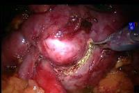
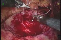
Tumor excision (left) and suture repair of defect (right)
The cancer is then removed with the margin of normal tissue, and placed in an impermeable specimen bag. Once the kidney cancer has been removed, the kidney is repaired by suture closing the collecting system (plumbing within the kidney) and then by closing the functioning kidney tissues that makes up the majority of the kidney. The repair of the area of the kidney where the kidney cancer has been removed is known as reconstruction of the kidney. After the kidney has been reconstructed, the blood supply to the kidney is restored, and the specimen bag is removed from the body from one of the small incisions. While the patient is still under anesthesia, the surgeon may work with a pathologist who microscopically examines the specimen further. Once complete removal of the kidney cancer has been confirmed, a small tube may be left in the area of the surgery (drain) to remove any excess fluids that may collect in the area.
Recovery
Once the surgery is complete, the patient is transferred to the recovery room for observation and then to a hospital room. Typically, on the day of surgery, the patient can drink liquids, and may be able to do some walking, relatively comfortably with limited pain. On the day after surgery it is common to have a regular diet. If everything has gone well, most people are discharged home on the first or second day after surgery with limited well-controlled pain, eating a regular diet, and feeling well. On discharge, oral antibiotics and oral pain medications are typically prescribed, and a follow-up office visit is scheduled for a week later. While patients will typically be able to do routine activities such as eating and taking care of everyday needs, it is generally suggested that no heavy lifting or vigorous activity be performed for at least four weeks to allow the body to recover. While it is surgeon dependent, most surgeons will suggest only light lifting (less than 10lbs), gentle activity, and no driving for one to two weeks after surgery. It is typical for patients to feel drained or lack energy for several weeks after surgery, and complete and full return to activity will usually take four to six weeks. However, every patient is different and recovery is somewhat variable. Generally, the patient will feel better day by day.
Advantages/Disadvantages
da Vinci robotic laparoscopic partial nephrectomy has the advantages intrinsic to any procedure in which the cancer is completely removed. With the da Vinci robotic laparoscopic partial nephrectomy, patients return to full activity in less than half the time it takes to recover from open partial nephrectomy. However, as it is a very technically challenging operation, it is not offered at many medical centers. Drs. Trapasso and Maggioncalda have created a highly trained, dedicated team of surgeons, anesthesia personnel, and operating room staff at Lehigh Valley Hospital to be able to offer this procedure to their patients. Overall, this robotic technology is a remarkable adjunct to the experienced surgeon who is familiar with kidney cancer, and laparoscopic anatomy and technique.