What is a CT Scan of the Body?
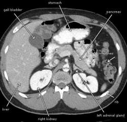
A CT scan—sometimes called CAT scanning—is a noninvasive medical test that helps physicians diagnose and treat medical conditions. It combines special x-ray equipment with sophisticated computers to produce multiple images or pictures of the inside of the body. These cross-sectional images of the body area being studied can then be examined on a computer monitor, printed and transferred to a compact disc. It's one of the best and fastest tools for studying the chest, abdomen and pelvis because it provides detailed, cross-sectional views of all types of tissue. This technology is often the preferred method for diagnosing kidney stones, and also many different cancers. The images allow a physician to not only detect the presence of a tumor but also measure its size, precise location and the extent of the tumor's involvement with other nearby structures.
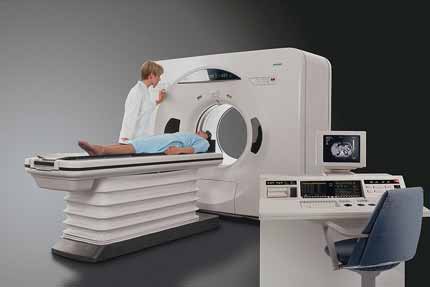
The CT scanner is typically a large, box like machine with a hole, or short tunnel, in the center. You will lie on a narrow examination table that slides into and out of this tunnel. Rotating around you, the x-ray tube and electronic x-ray detectors are located opposite each other in a ring, called a gantry. CT scanning of the body is usually completed within 15 minutes. The computer workstation that processes the imaging information is located in a separate room, where the technologist operates the scanner and monitors your examination.

You should wear comfortable, loose-fitting clothing to your exam. You may be given a gown to wear during the procedure. Metal objects including jewelry, eyeglasses, dentures and hairpins may affect the CT images and should be left at home or removed prior to your exam. You may also be asked to remove hearing aids and removable dental work.
Also, inform your doctor of any recent illnesses or other medical conditions, and if you have a history of heart disease, asthma, diabetes, kidney disease or thyroid problems. Women should always inform their physician and the CT technologist if there is any possibility they may be pregnant.
A physician, usually a radiologist with expertise in supervising and interpreting radiology examinations, will analyze the images and send a signed report to your urologist or the physician who referred you for the exam, who will then discuss the results with you.
Benefits
- CT scanning is painless, noninvasive and accurate.
- A major advantage of CT is its ability to image tumors, stones, bone, soft tissue and blood vessels all at the same time.
- Unlike conventional x-rays, CT scanning provides very detailed images of many types of tissue as well as the lungs, bones, and blood vessels.
- CT examinations are fast and simple; in emergency cases, they can reveal internal injuries and bleeding quickly enough to help save lives.
- CT has been shown to be a cost-effective imaging tool for a wide range of clinical problems.
- CT is less sensitive to patient movement than MRI.
- CT can be performed if you have an implanted medical device of any kind, unlike MRI
- A diagnosis determined by CT scanning may eliminate the need for exploratory surgery and surgical biopsy.
- No radiation remains in a patient's body after a CT examination.
- X-rays used in CT scans usually have no immediate side effects.
Risks
- There is always a slight chance of cancer from excessive exposure to radiation. However, the benefit of an accurate diagnosis far outweighs the risk.
- The effective radiation dose from this procedure ranges from approximately two to 10 mSv, which is about the same as the average person receives from background radiation in to 8 months to three years.
- Women should always inform their physician and x-ray or CT technologist if there is any possibility that they are pregnant.
- CT scanning is, in general, not recommended for pregnant women unless medically necessary because of potential risk to the baby.
Low Dose Radiation Exposure CT Scan
Recent reports have suggested that radiation doses from CT scans may place patients at an increased risk of future cancer. On an individual basis, it's typically not a substantial risk, but still a risk applied to an increasingly large population of those who are obtaining repeated scans. At USLV, we have incorporated new software technology that can significantly reduce the radiation dose of certain CT scan studies. This software works as a filter that reduces random noise and enhances visibility of structures needing to be examined. In this way, it is possible for us to obtain high-quality images, while at the same time lowering the dose by as much as 70 percent, and thereby reducing the radiation exposure to the patient as low as possible without compromising the quality of the test.

